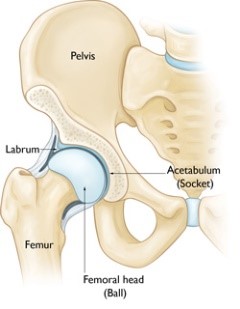
Hip arthroscopy is a surgical procedure that allows doctors to view the hip joint without making 2 or 3 small incisions (cut) through the skin and other soft tissues, a small camera, called an arthroscope, into your hip joint and the images are displayed on a monitor. Your surgeon uses these images to guide miniature surgical instruments. This results in less pain for patients, less joint stiffness, and often shortens the time it takes to recover and return to favourite activities as compared to an open (larger incision) surgery. Hip arthroscopy has been performed for many years, but is not as common as knee or shoulder arthroscopy.

HIP-ARTHROSCOPY
The hip is a ball-and-socket joint. The socket is formed by the acetabulum, which is part of the large pelvis bone. The ball is the femoral head, which is the upper end of the femur (thighbone). A smooth cushion-like tissue known as the articular cartilage covers the surface of the ball and the socket creating a smooth, low friction surface that helps the bones glide easily across each other during movement. The acetabulum is ringed by strong fibrocartilage called the labrum. The labrum forms a gasket around the socket, creating a tight seal and helping to provide stability to the joint. In FAI, bone overgrowth called bone spurs develop around the femoral head and/or along the acetabulum. This extra bone causes abnormal contact between the hip bones, and prevents them from moving smoothly during activity. Over time, this can result in tears of the labrum and breakdown of articular cartilage (osteoarthritis). Hip arthroscopy may relieve painful symptoms of many problems that damage the labrum, articular cartilage, or other soft tissues surrounding the joint. Your treating orthopaedic Surgeon may recommend hip arthroscopy if you have a painful condition that does not respond to nonsurgical treatment such as rest, physical therapy, and medications or injections that can reduce inflammation.
Conditions can be treated with Hip Arthroscopy?
- Femoro-acetabular impingement (FAI)
- Labral tears/Dysplasia
- Snapping hip syndromes
- Synovitis
- Loose bodies
- Hip joint infection (rarely)
Preparing for Surgery
Your treating surgeon may ask you to schedule a complete physical examination with your physi-cian several weeks before the operation. This is needed to make sure you are healthy enough to have the surgery and complete the recovery process.
- Tests- Several tests, such as blood and urine samples may be needed to help your surgeon plan your surgery.
- Medications- Your treating surgeon will ask you to inform all the medications that you take as it is im-portant to stop certain medications for few days/weeks prior to undergoing a surgery.
- Urinary Evaluations- History of recent/frequent urinary infections and prostrate disease in men mandates a thorough evaluation before surgery.
- Social Planning - You will be able to walk on crutches/walker soon after surgery. Although you should be independent soon after surgery, depending on your recovery, you may need help/assistance for a few days.
The Hospital Stay
- Admission- You will most likely be admitted to the hospital one night prior to your surgery.
- Anesthesia- After admission, you will be evaluated by a member of the anaesthesia team. The most common types of anaesthesia are general anaesthesia (you are put to sleep) or spinal, epidural, or regional nerve block anaesthesia (you are awake but your body is numb from the waist down). The anaesthesia team, with your input, will determine which type of anaesthesia will be best for you.
- Procedure- The procedure itself takes approximately 2 - 3 hours. Your orthopaedic surgeon will first assess the damage within your hip joint through an arthroscope and then make further small incisions to introduce instruments to repair the damage.
- Post-Surgery- After surgery, you will be moved to the recovery room, where you will remain for sev-eral hours while your recovery from anaesthesia is monitored. After you wake up, you will be taken to your hospital room.
- Post–Operative Period- You will most likely stay in the hospital for 2 - 3 days. During this time you will be under supervision of the expert panel of the hospital involved in your treatment. You will be en-couraged to mobilise by the physiotherapy team using walking aids.
The Procedure
- Positioning-At the start of the procedure, your leg will be put in traction and your hip will be pulled away from the socket enough to insert instruments, see the entire joint, and perform the treatments needed.
- Procedure-After traction is applied, a small puncture incision is made in your hip for the arthroscope. Through the arthroscope your surgeon can view the inside of your hip and identify dam-age. Images from the arthroscope are projected on the video screen showing your surgeon the inside of your hip and any problems. Once the problem is clearly identified, other small instruments are inserted through separate incisions to repair it. At the end of sur-gery, the arthroscopy incisions are usually stitched or covered with skin tapes. An absor-bent dressing is applied to the hip.
Are there any complications of Hip Arthroscopy
Complications from hip arthroscopy are uncommon. As with any other surgery, some known complications although extremely rare that you should be aware of include-
- Injury to the surrounding nerves or blood vessels, or the joint itself.
- Traction needed for the procedure can stretch nerves and cause numbness, but this is usu-ally temporary.
- Infection
- Blood clots or deep vein thrombosis
- Pain Management- After surgery, you will feel some pain. This is a natural part of the healing process. Your doctor and nurses will work to reduce your pain. Many types of medicines are available to help manage pain, including opioids, non-steroidal anti-inflammatory drugs (NSAIDs), and local anaesthetics. Your doctor may use a combination of these medications to im-prove pain relief, as well as minimise the need for opioids.
- Blood Clot Prevention - Your orthopaedic surgeon may prescribe one or more measures to prevent blood clots and decrease leg swelling. These may include special support hose, inflatable leg cov-erings (compression boots), and blood thinners. Foot and ankle movement also is en-couraged immediately following surgery to increase blood flow in your leg muscles to help prevent leg swelling and blood clots.
- Physical Therapy- Most patients begin exercising the day after surgery. A physical therapist will teach you specific exercises to strengthen your leg and restore hip movement to allow walk-ing and other normal daily activities soon after your surgery
- Bearing Weight - Crutches may be necessary after your procedure. In some cases, they are needed only until any limping has stopped. If you required a more extensive procedure, however, you may need crutches for 1 to 2 months
- Rehabilitation Exercise- Your surgeon will develop a rehabilitation plan in consultation with your physiothera-pist based on the surgical procedures you required. Specific exercises to restore your strength and mobility are important but not essential
Some Key issues
If you have any question concerning your surgery is risk, benefits, likely outcome or complication please do not hesitate to contact a team member at Joint & Sports Clinic.
